Describe How a Light Microscope Creates a Magnified Image
Quality magnified real image. So a lens magnifying ten times would be 10.

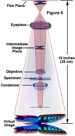
Anatomy Of The Microscope The Concept Of Magnification Olympus Ls
How lenses magnify When you look through a simple light microscope or a magnifying glass you are looking through a biconvex lens one thats bent like the back of a spoon on both sides made of glass.

. When the microscope is properly illuminated both the object and the edges of the field aperture diaphragm should be in the same plane of focus and the field iris diaphragm should be centered in the field of view. The image is then magnified by a second lens called an ocular lens or eyepiece as it is brought to your eye. Electron beams are used in place of visible light to produce the magnified image.
Zdescribe the principle of light microscope zexplain the parts of a light microscope. An objective lens and an eyepiece. This light is called direct undeviated or non-diffracted light and represents the background light.
There are two types TEM and SEM. A microscope must be able not only to magnify objects sufficiently but also to resolve or separate the fine details of the object that are of interest to the viewer. The human eye can perceive changes in light amplitude intensity.
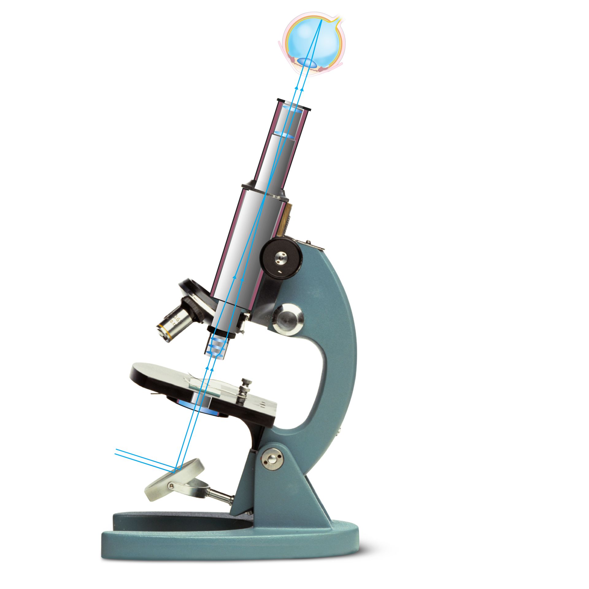
The image is then passed through one or two lenses for magnification for viewing. We get around the problems of brightness and depth of field by using something besides light for the microscope. In the optical microscope visible light rays reflected from or transmitted by the viewed object pass through a series of lenses and form an enlarged image of the object.
In the optical microsco Unlock 15 answers now and every day. How the Microscope Forms Images. It uses light to project a magnified view of the object.
A light microscope creates a magnified image through a series of lenses. The functioning of the light microscope is based on its ability to focus a beam of light through a specimen which is very small and transparent to produce an image. Most light microscopes are compound microscope that contains at least two lenses.
There are two lenses. How much the specimen is enlarged depends on the thickness of the lens how curved the lens is and how fast light moves through the object being viewed. A light microscope uses incident light and a sequence of lenses to create a magnified image.
In the optical microscope when light from an illumination source passes through the condenser and then through the specimen some of the light passes both around and through the specimen undisturbed in its path. 452014 42527 PM. Transmission electron microscope Electrons pass through the specimen and are scattered.
The third type of lens called the condenser lens focuses the light rays and then the objective lens and eyepiece magnify the specimen. The more you magnify an image the thinner the light gets spread and you reach the point where even with a very bright light the image is too dark to see anything. Body tube Body tube can be monocular binocular and the combine photo-binocular also.
An electromagnetic lens focuses the image onto a fluorescent screen or photographic plate. A light microscope uses a beam of light that normally pass through the lenses to produce an image of a certain object or a specimen being viewed. The magnification of a lens is shown by a multiplication sign followed by the amount the lens magnifies.
The light rays which reflect off of the object are then focused into a magnified image. The light rays reflected from the viewed abject pass through these many lenses and form an enlarged picture of the object. Magnifying powers of objectives are from 11 to 1001.
This type of microscopy is a Phase-Contrast using two light beams passing through prisms. The total magnification of. It brings the image of the object into focus at a short distance within the microscopes tube.
This makes the light spread out after it passes through the lens producing an enlarged and inverted upside down and backwards image of the specimen. Here are a number of highest rated Light Microscope pictures on internet. Rather than focusing light at a specimen electron microscopes utilize streams of electrons which are accelerated in a vacuum and directed at a prepared specimen.
A light microscope creates a magnified image through a series of lenses. We consent this nice of Light Microscope graphic could possibly be the most trending subject afterward we part it in google gain or facebook. The object being viewed is on the far side of the lens.
Its submitted by government in the best field. The other major difference between a telescope and a microscope is that a microscope has a light source and a condenser. A microscope must be able not only to magnify objects sufficiently but also to resolve or separate the fine details of the object that are of interest to the viewer.
We identified it from trustworthy source. Images like the one below are taken with a scanning electron. A light microscope focuses a light source at a specimen through a series of lenses.
Step-by-step explanation Light microscope magnification increase while its resolution decrease. A light microscope focuses a light source at a specimen through a series of lenses. The transparency of the specimen allows easy and quick penetration of light.
A microscope is an instrument that produces a clear magnified image of an object viewed through it. Parts of a Light Microscope.

How Does An Optical Microscope Work Dk Find Out

Light Microscopes An Overview Sciencedirect Topics

Molecular Expressions Microscopy Primer Anatomy Of The Microscope Magnification
No comments for "Describe How a Light Microscope Creates a Magnified Image"
Post a Comment